
IGCSE Biology: Coordination and Response
Complete Cambridge IGCSE Biology guide covering the nervous system, reflex arcs, neurones, synapses, the human eye, accommodation, pupil reflex and adrenaline.
Learning Objectives
Define coordination and explain why organisms need to respond to stimuli.
Describe receptors, effectors and the nervous system.
Explain the pathway of a reflex arc.
Compare sensory, relay and motor neurones.
Describe how impulses cross synapses.
Explain accommodation, pupil reflex and the effects of adrenaline.
Human Nervous System
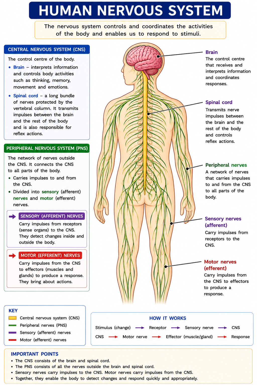
The nervous system allows the body to detect stimuli and respond quickly. It is made of the central nervous system and peripheral nerves.
| Part | Description |
|---|---|
| Central nervous system | The brain and spinal cord. |
| Peripheral nervous system | Nerves outside the brain and spinal cord. |
| Sensory neurones | Carry impulses from receptors to the CNS. |
| Motor neurones | Carry impulses from the CNS to effectors. |
Receptors, Effectors and Stimuli
| Term | Meaning | Example |
|---|---|---|
| Stimulus | A change in the environment. | Light, heat, sound, pressure. |
| Receptor | A cell or organ that detects a stimulus. | Retina in the eye, skin receptors. |
| Effector | A muscle or gland that produces a response. | Muscle contracts, gland secretes hormone. |
| Response | The action caused by the effector. | Hand pulls away from a hot object. |
Reflex Arc
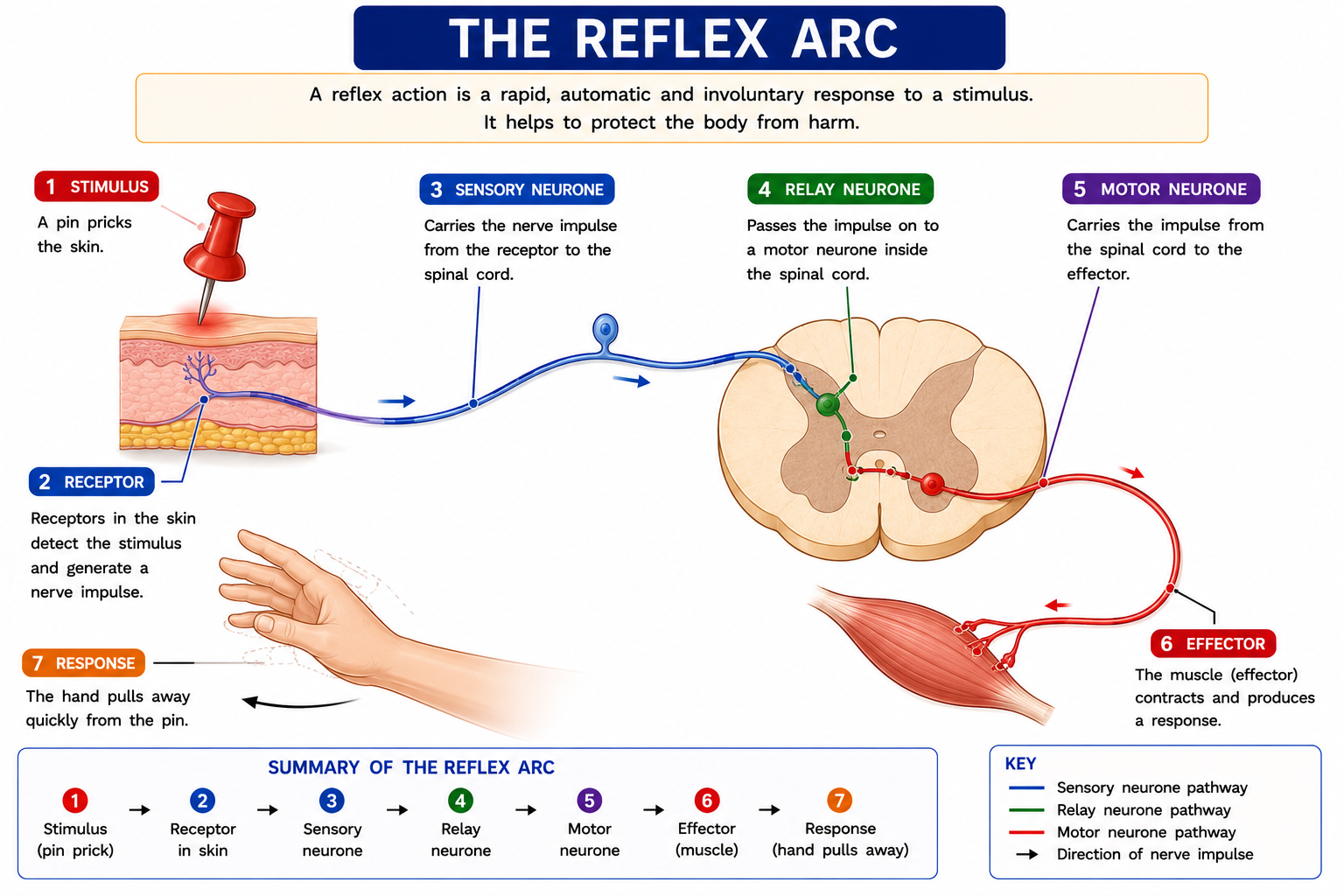
A reflex action is rapid, automatic and involuntary. It protects the body from harm.
Pathway: stimulus → receptor → sensory neurone → relay neurone → motor neurone → effector → response.
Types of Neurones
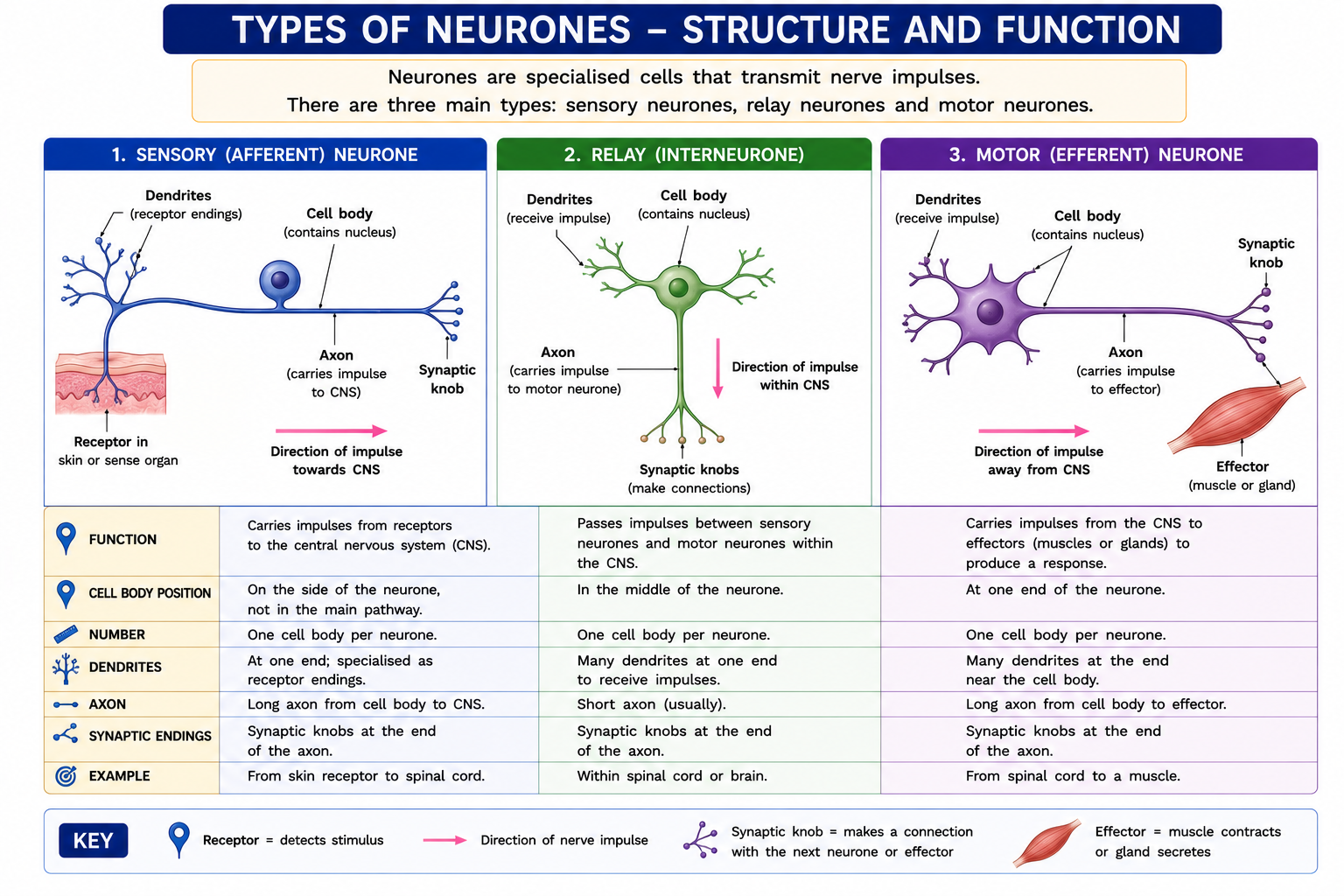
| Feature | Sensory neurone | Relay neurone | Motor neurone |
|---|---|---|---|
| Function | Carries impulses from receptors to CNS. | Passes impulses within CNS. | Carries impulses from CNS to effectors. |
| Location | Between receptor and CNS. | Brain or spinal cord. | Between CNS and effector. |
| Direction | Towards CNS. | Within CNS. | Away from CNS. |
Synapse
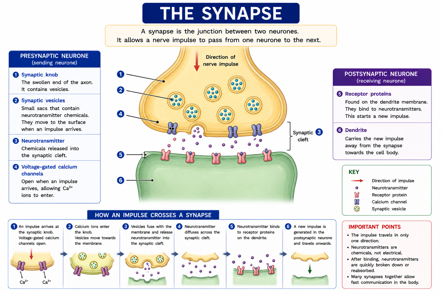
A synapse is a junction between two neurones. Electrical impulses cannot cross the gap directly, so chemicals called neurotransmitters diffuse across the synaptic cleft.
- An impulse reaches the synaptic knob.
- Neurotransmitter is released into the synaptic cleft.
- Neurotransmitter diffuses across the gap.
- It binds to receptors on the next neurone.
- A new impulse is started in the next neurone.
Human Eye Structure

| Part | Function |
|---|---|
| Cornea | Refracts light entering the eye. |
| Iris | Controls the size of the pupil. |
| Pupil | Allows light to enter the eye. |
| Lens | Focuses light onto the retina. |
| Retina | Contains light-sensitive cells. |
| Fovea | Area of sharpest vision. |
| Blind spot | Where the optic nerve leaves the eye; no light receptors. |
| Optic nerve | Carries impulses from the retina to the brain. |
Accommodation
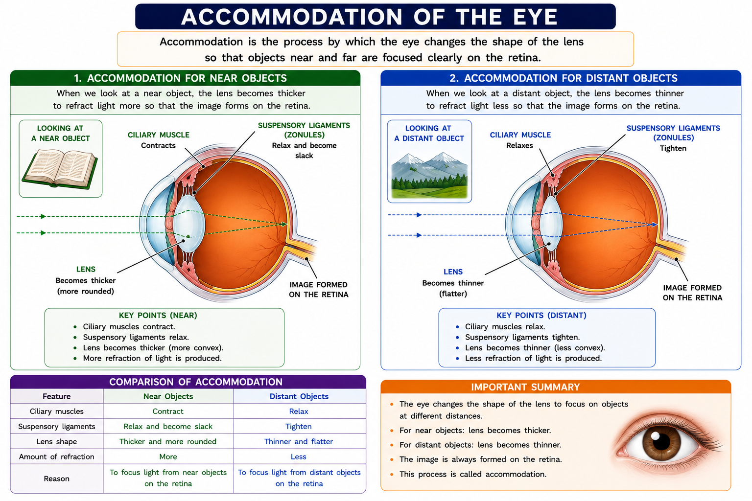
| Feature | Near Object | Distant Object |
|---|---|---|
| Ciliary muscles | Contract | Relax |
| Suspensory ligaments | Relax | Tighten |
| Lens shape | Thicker / more rounded | Thinner / less rounded |
| Refraction | More refraction | Less refraction |
Pupil Reflex
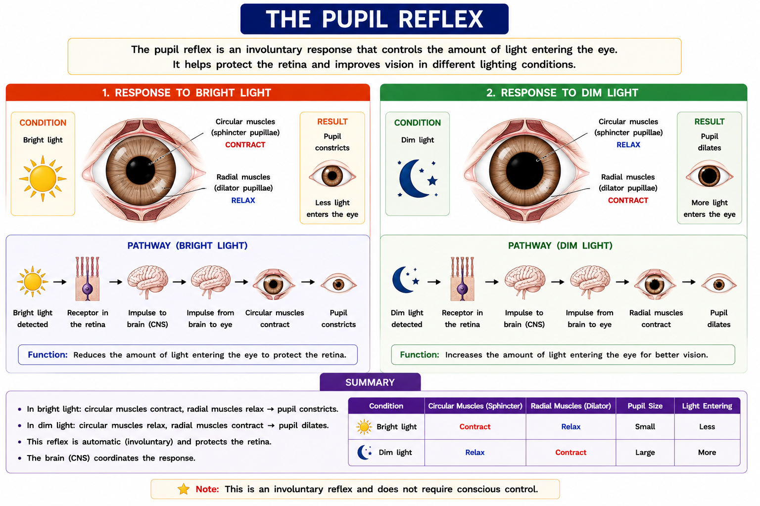
| Condition | Circular Muscles | Radial Muscles | Pupil | Effect |
|---|---|---|---|---|
| Bright light | Contract | Relax | Constricts | Less light enters to protect the retina. |
| Dim light | Relax | Contract | Dilates | More light enters for better vision. |
Hormones and Adrenaline
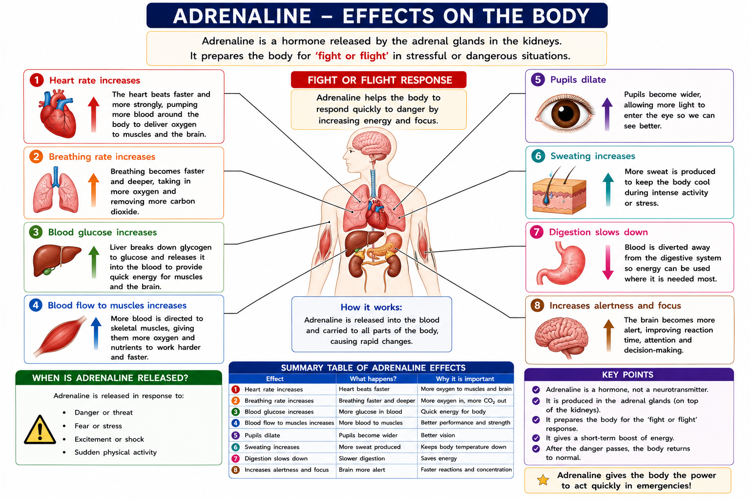
Adrenaline is a hormone secreted by the adrenal glands. It prepares the body for a fight-or-flight response.
| Effect of Adrenaline | Why It Helps |
|---|---|
| Increases heart rate | Delivers more oxygen and glucose to muscles. |
| Increases breathing rate | More oxygen enters the blood. |
| Increases blood glucose | Provides more glucose for respiration. |
| Redirects blood to muscles | Muscles can contract more strongly. |
| Dilates pupils | Allows more light into the eye. |
Exam Definitions
| Term | Definition |
|---|---|
| Coordination | The control of body activities so that responses are appropriate. |
| Stimulus | A change in the environment detected by receptors. |
| Receptor | A cell or organ that detects a stimulus. |
| Effector | A muscle or gland that produces a response. |
| Reflex action | A rapid, automatic and involuntary response to a stimulus. |
| Synapse | A junction between two neurones. |
| Accommodation | The process by which the lens changes shape to focus light from near or distant objects on the retina. |
| Hormone | A chemical substance produced by a gland and carried in the blood to a target organ. |
Common Exam Mistakes
- Confusing sensory neurones with motor neurones.
- Forgetting the relay neurone in the reflex arc.
- Saying reflex actions are conscious. They are involuntary.
- Writing that impulses cross synapses electrically only. Neurotransmitters are involved.
- Mixing up iris and pupil.
- Writing that the lens moves forward and backward. It changes shape.
- Reversing the roles of circular and radial muscles in the pupil reflex.
- Saying adrenaline is a neurotransmitter. In this chapter, it is treated as a hormone.
Exam Style Questions and Answers
1. Define a stimulus.
A stimulus is a change in the environment detected by receptors.
2. State the pathway of a reflex arc.
Stimulus → receptor → sensory neurone → relay neurone → motor neurone → effector → response.
3. Why are reflex actions important?
They are rapid, automatic responses that protect the body from harm.
4. What is the function of a synapse?
A synapse allows an impulse to pass from one neurone to another using neurotransmitters.
5. What is the function of the retina?
The retina contains light-sensitive cells that detect light and start nerve impulses.
6. Describe accommodation for a near object.
Ciliary muscles contract, suspensory ligaments relax and the lens becomes thicker to refract light more.
7. What happens to the pupil in bright light?
Circular muscles contract, radial muscles relax and the pupil constricts.
8. Give two effects of adrenaline.
Adrenaline increases heart rate and breathing rate. It also increases blood glucose and blood flow to muscles.
Quick Revision Summary
- The nervous system coordinates fast responses.
- The CNS consists of the brain and spinal cord.
- Sensory neurones carry impulses to the CNS.
- Motor neurones carry impulses from the CNS to effectors.
- Reflex actions are rapid, automatic and involuntary.
- Synapses use neurotransmitters to pass impulses between neurones.
- The lens changes shape during accommodation.
- Bright light causes the pupil to constrict.
- Dim light causes the pupil to dilate.
- Adrenaline prepares the body for fight or flight.